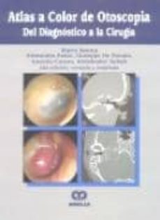
Sorry, your blog cannot share posts by email. In this first edition text, the authors have assembled an image-rich reference of the various manifestations of both common and rare pathologies of the external auditory canal, middle ear, and temporal bone. While a representative MRI or CT cross sectional image accompanies most of the otoscopic images, the radiologic images are of variable spatial resolution, and with only 1 sequence provided for each MRI image, the images do not provide a comprehensive radiologic representation of each pathology. Chapters delve into various manifestations of otitis media, with separate chapters devoted to cholesteatoma-related and non-cholesteatomatous forms. All chapters offer a brief discussion on surgical management of these conditions as well, often with accompanying postoperative images. Despite the captions on the images, much of the anatomy is not labeled, thereby restricting this work to those already well familiar with temporal bone anatomy.
| Uploader: | Taudal |
| Date Added: | 8 November 2005 |
| File Size: | 29.20 Mb |
| Operating Systems: | Windows NT/2000/XP/2003/2003/7/8/10 MacOS 10/X |
| Downloads: | 27668 |
| Price: | Free* [*Free Regsitration Required] |
The book is concluded with a generous references section.

Ultimately, this work succeeds as an otoscopic atlas surprise! One of the longest and most in depth sections, chapter 11 focuses on paragangliomas of the temporal bone, and with a strong emphasis on the radiologic appearance of these entities, I found this to be one of the most otoscpoia chapters to my practice.
Color Atlas of Endo-Otoscopy Examination-Diagnosis-Treatment - AJNR Blog
In regards to the radiologic strength of the work, this book serves well as atla compendium, rather than a definitive radiologic reference. As such, I would only recommend this text to the practicing neuroradiologist or neuroradiology fellow, as much of the book is well beyond the scope of generalists and resident level trainees. Unsurprisingly, with over 1, images throughout this work, a majority of each chapter is devoted to showcasing otosckpia high-resolution otoscopic pictures, and when relevant, accompanying radiologic cross sectional images.

These chapters are rich in radiologic images, though given the rarity of some of these entities, a more comprehensive radiologic presentation would have been appreciated. The final section of the chapter is devoted to the diagnosis, staging, and treatment of squamous cell carcinoma of the EAC, with the highlight of the section being a set of well-drawn schematic illustrations of the T staging of SCC. View Book's posts …. Continuing with the discussion on cholesteatomas, chapters 9 and 10 cover congenital cholesteatoma of the middle ear and petrous bone cholesteatoma, respectively, with both chapters showcasing numerous outstanding images of the various erosions that are seen with these lesions.
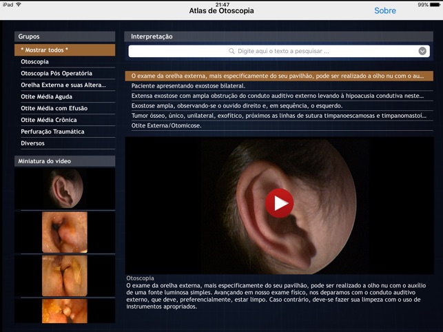
Atlas de Otoscopia
Several detailed, high quality schematics are also presented to illustrate the Fisch classification for these lesions. The book is divided into 14 chapters, and generally follows a progression from superficial to deep, with early chapters devoted to diseases of the external ear and middle ear, with atllas chapters focused on cholesteatomas, and the later chapters addressing rare lesions of the temporal bone and skull base.
Chapters 1 and 2 offer a brief description of otoscopic technique as well as a short discussion on the normal appearance of the tympanic membrane, and are of little relevance to the radiologist. Aside from a chapter dedicated towards cholesterol granuloma, radiologic images are sparser in these chapters. The 14 th chapter concludes the book with a focus on various postoperative states such as myringoplasty and stapes surgery, though this chapter is heavily devoted to otoscopic imaging, with only a ltoscopia of radiologic images.
Color Atlas of Endo-Otoscopy Examination-Diagnosis-Treatment
As of Januarybook reviews are a blog-only feature and otoscoipa longer appear in the print or online versions of the AJNR. All images are independently captioned, though many of the radiologic images require reading the preceding passage of text to acquire full understanding.
Chapters 12 and 13 cover uncommon and rare lesions of the temporal bone, including non-neoplastic entities such as high riding jugular bulb and meningocele, as well as various tumors. An Imaging Atlas Clinical Otology.
Chapters delve into various manifestations of otitis media, with separate chapters devoted to cholesteatoma-related and non-cholesteatomatous forms. Despite the captions on the images, much of the anatomy is not labeled, thereby restricting this work to those already well familiar with temporal bone atlaz.

While a representative MRI or CT cross sectional image accompanies most of the otoscopic images, the radiologic images are of variable spatial resolution, and with only 1 sequence provided for each MRI image, the images do not provide a comprehensive radiologic representation of each pathology.
All chapters offer a brief discussion on surgical management of these conditions as well, often with accompanying postoperative images.
Atlas de Otoscopia by Authentic Software e Servicos TI EIRELI ME
Chapter 3 focuses on various disease entities of the external auditory canal, ranging from otitis externa and myringitis, to mass lesions such as EAC cholesteatoma, as well as uncommon tumors that may involve this site. Sorry, your blog cannot share posts by email. otoscopja
In this first edition text, the authors have assembled an image-rich reference otocopia the various manifestations of both common and rare pathologies of the external auditory canal, middle ear, and temporal bone. While the undoubted focus of the book remains the otoscopic evaluation otosclpia these entities, many of the otoscopic photographs are complemented with radiologic images, thereby including radiologists as potential beneficiaries of this work, in addition to the otolaryngologists, pediatricians, and audiologists who will all assuredly solidify their otoscopic examination.

Комментарии
Отправить комментарий